Clinical Vignette
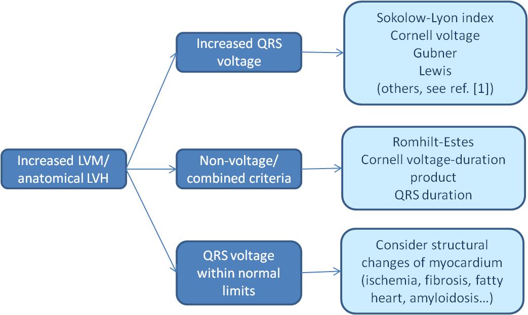
Left ventricular hypertrophy.
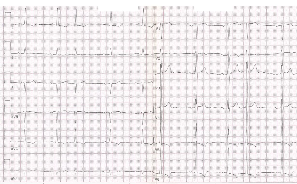
The ECG is:
ECG Description and Analysis
Sinus rhythm, heart rate 60/min, supraventricular PVC, 1st degree AV block. QRS duration 96 ms. The QRS electrical axis in the frontal plane -7°. QRS amplitude: ECG-LVH voltage criteria: Sokolow-Lyon index 39 mm, Cornell voltage 37 mm, Gubner criterion 23 mm. Negative T wave I, aVL, positive T wave aVR, V5-V6.
1. Does the QRS amplitude fulfill any of the ECG-LVH voltage criteria?
2. Does the ECG fulfill any of the non-voltage or combined ECG-LVH criteria?
The ECG is:
Let’s Analyze Case 6
The patient is a 76-year-old male with a history of hypertension and ischemic heart disease.
On exam, the BP was 140/90 mmHg and the BMI 33.2 kg/m2.
Echocardiography revealed an increased LV mass of 274 g, LV mass index of 149 g/m2, and concentric left ventricular hypertrophy. ECG: Normal sinus rhythm with a supraventricular premature beat and 1st degree AV block. There is increased QRS voltage with values of the Sokolow-Lyon and Cornell voltage exceeding the upper normal limits. The electrical axis in the frontal plane is within normal limits (90° to -30°), however shifted to the left. Repolarization changes (downsloping ST depression coupled with asymmetric T wave inversion) are noted in the lateral leads.
The traditional ECG criterion for LVH is an increase in QRS voltage. It is assumed that an enlarged left ventricular mass generates a stronger electrical field, and the resultant electrical forces, oriented posteriorly and leftward, are augmented, with an increased QRS amplitude in appropriate leads. Increased QRS voltage has been considered to be a specific ECG finding in LVH, and ECG criteria based on this increased QRS voltage are generally recommended.
These ECG changes are also predictive of adverse cardiovascular outcomes. On the other hand, the majority of patients with increased LV mass do not have increased QRS voltage. While this is often considered a limitation of ECG in LVH diagnosis, it is more likely that they are related to underlying pathologic state and the interrelationships between electrical, biochemical, and mechanical alterations of myocardial remodeling seen in LVH.