Clinical Vignette
Two cases of acute Anterior STEMI. The importance of knowing the site of occlusion.
The ECGs are:
ECG Description and Analysis
Questions that we must ask:
1. What ECG shows an area of ischemia more extensive?
2. How do I know that the bottom ECG does not correspond to the RCA or LCX occlusion?
3. Where is the occlusion located in both ECGs?
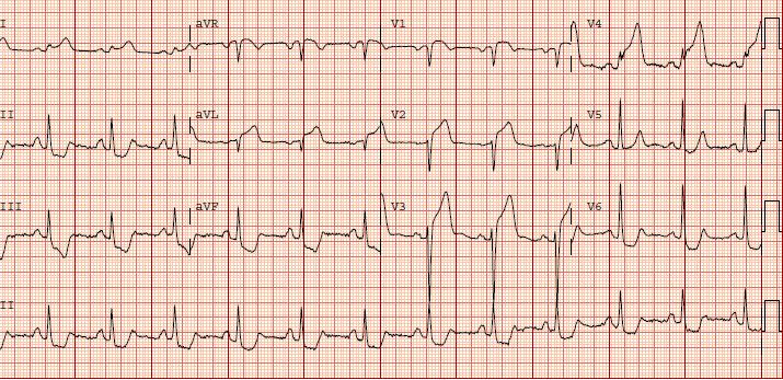
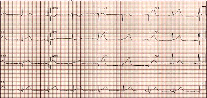
The ECGs are:
Let’s Analyze Case 3
What ECG shows an area of ischemia more extensive?
The top ECG is a typical case of STEMI due to proximal LAD occlusion before the first diagonal and first septal branches.
[Detailed analysis continues with images and explanations.]
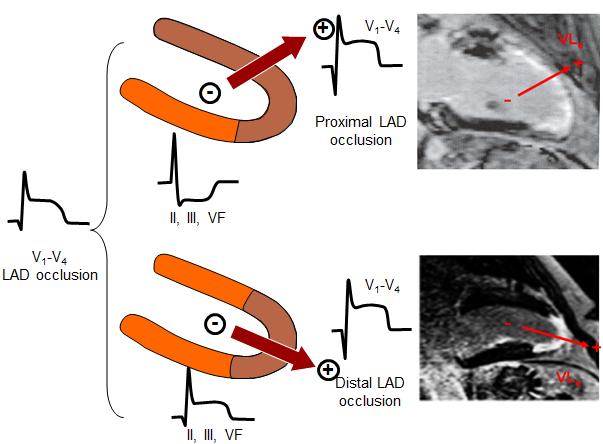
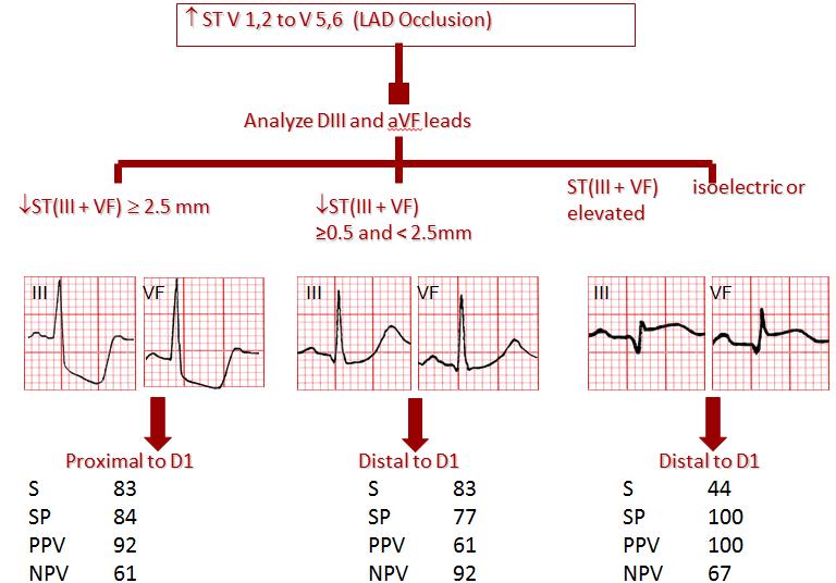
Where is the occlusion located in both ECGs?
The figure below shows the algorithm to locate the zone of LAD occlusion in case of STEMI with predominant ST-segment elevation in precordial leads.