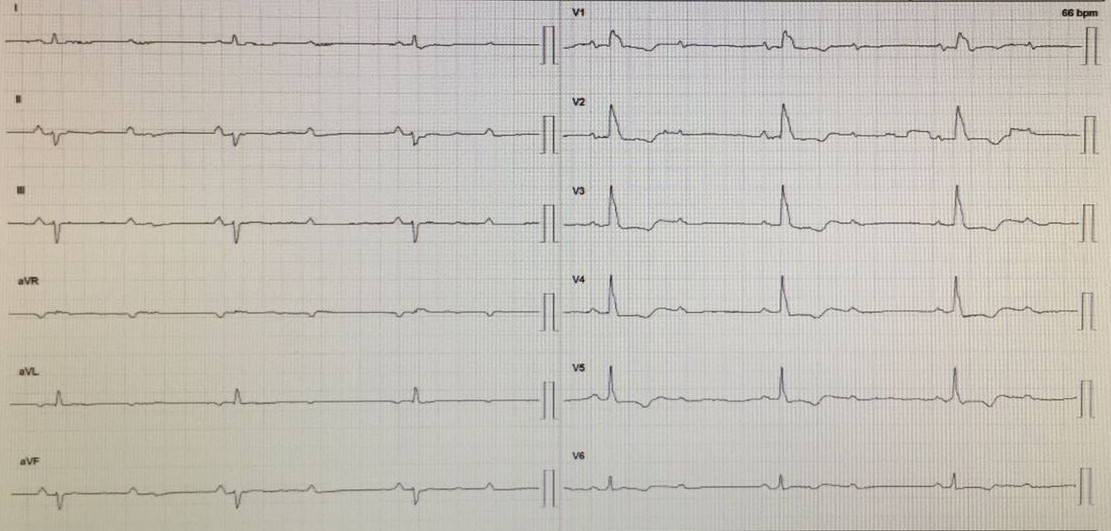
Clinical Vignette
A 62 y-o-w was referred for dizziness and one episode of syncope while watching TV. She has a previous AMI two years ago with a negative stress test three months ago.
The ECG is:
ECG Description and Analysis
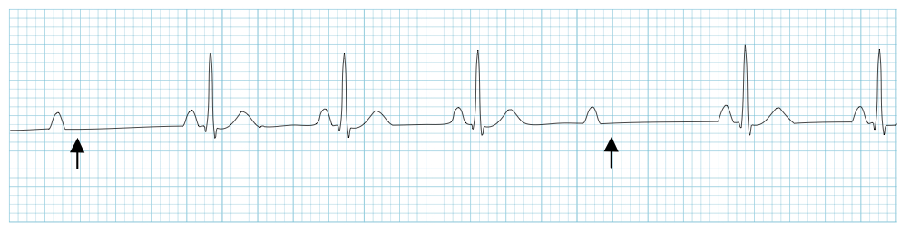
1. Are there missing QRS?
2. P-P interval are regular or irregular?
3. Are the PR intervals stable or increasing?
Let’s Analyze Case 1
This is a second degree AV block where some but not all the P waves are conducted.
Arrows at the strip show the missing QRS.
There are 2 types of 2nd degree AV block, Mobitz I with Wenckebach periods and Mobitz II without them.
In this ECG there is no increasing of the PR interval.
Mobitz I is usually due to a functional suppression of AV conduction (e.g. due to drugs, reversible ischaemia)
while Mobitz II is usually due to failure of conduction at the level of the His-Purkinje system (i.e. below the AV node),
is also more likely to be secondary to structural damage to the conducting system (e.g. infarction, fibrosis, necrosis)
and the risk of asystole per year is high.
In the case of 2:1 block (in the ECG, 2 P waves for every QRS complex) it is impossible to differentiate type I from type II Mobitz block
based solely on the P:QRS ratio or on a pattern of lengthening PR intervals.
In this case, a normal PR interval with a widened QRS is most likely indicative of a type II-like pathology.
On the other hand, a lengthened PR interval with a normal QRS width is most likely indicative of a type I AV block.
Type II Mobitz block usually occurs with a fixed P:QRS ratio, with a set number of P waves for every successfully conducted QRS.
This ratio is also frequently specified in referring to "3:1", "4:1", "5:1", or higher Mobitz type II block.
Higher numbers of P waves for every QRS indicate more severe block.
Remember the difference between Type 1 and Type 2 second degree heart block:
• Type 1 has increasing PR intervals, increasing until the QRS is missing
• Type 2 has constant PR intervals, with randomly dropped QRS complexes
Second degree type 2 heart blocks usually occur BELOW the AV node:
• 20% are in the Bundle of His
• 80% are in the bundle branches (note that both branches would need to be blocked at the same time)
Exercise Based on Case 1
1. Use your calipers to determine the exact heart rate and P-P interval.
2. Use your calipers to determine the PR interval. Are they equal?
3. Review all possible differences with second degree AV block with Wenckebach periods.
What’s Your Diagnosis?
2nd degree AV block type II (Mobitz)